%20(3).png?width=1555&height=462&name=Novarad%20EHS%20Logo%20Full%20Color%20(1)%20(3).png "Novarad Enterprise Healthcare Solutions")
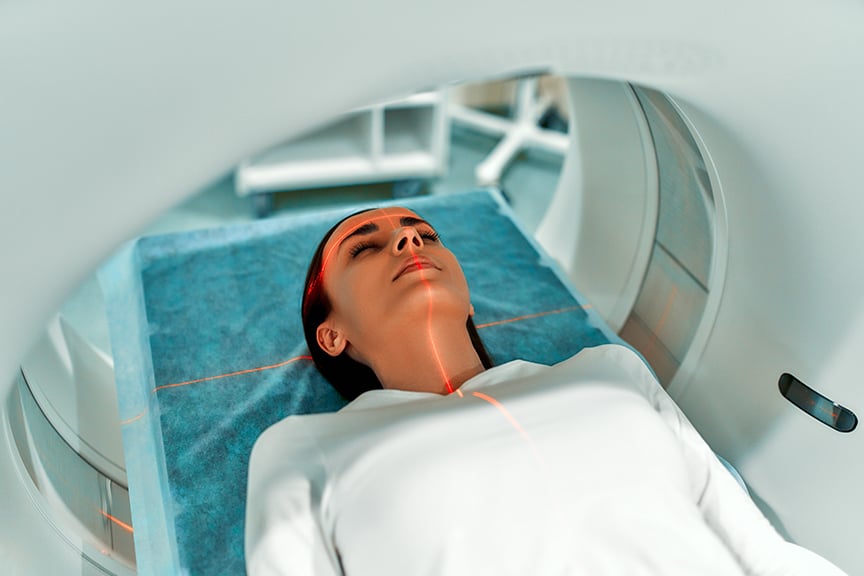
The increasing use of radiological imaging in recent years has caused doctors and healthcare workers to ask how they can reduce radiation exposure from X-Rays, CT scans, and other imaging equipment. In 2010, a large-scale study determined that an estimated 70 million CT scans are performed in the U.S. every year. This is an enormous increase from the 3 million per year in the 1980s. As a result, as many as 14,000 people may die every year of radiation-induced cancers.
X-rays and CT scans have become a necessary medical tool with a host of miraculous benefits. Despite these benefits, the risk to both doctors and patients has been acknowledged by the FDA, and safety measures are being taken to decrease exposure.
Due to the nature of their work, doctors, healthcare workers, and hospital staff face increased danger of exposure, making it crucial to have policies and procedures in place to minimize these risks. In this article, we discuss the dangers of long-term exposure to low dose ionizing radiation, as well as methods for medical professionals to minimize dosage so they can continue using these life-saving tools safely and effectively.
What are the Dangers of Ionizing Radiation?
We are all exposed to electromagnetic radiation in varying forms and degrees throughout our lifetimes. Radiation can be ionizing or non-ionizing depending on its frequency. Ionizing radiation is on the high-frequency end of the electromagnetic spectrum, and we all absorb this type of radiation in very small doses from natural sources. These sources include soil, water, vegetation, and the sun. Receiving natural radiation here and there throughout our lives is not generally dangerous, but small doses from man-made sources can add up.
X-rays and gamma rays are forms of ionizing radiation, or high-frequency energy. Both X-rays and CTs use X-rays. To produce an image, these high-energy photons pass through the body and onto a detector. As they do so, they deposit energy into the body. X-ray's high-frequency ionizing radiation has enough energy to remove an electron from (ionize) an atom or molecule. This ionization can damage DNA and possibly lead to cancer later in life.
Radiation dosage is cumulative. Exposure to one CT scan or X-ray is unlikely to cause cancer later in life, but this risk increases with each scan. This is why it is important that doctors make it top priority to minimize their exposure.
Studies have shown that a radiation dose of 10 mSv (millisieverts) is equivalent to a 1 in 2000 risk of developing fatal cancer later in life. According to Harvard Health, most X-ray scans range from 0.001 to 1.5 mSv, and most CT scans range from 2 to 16 mSv. So again, being exposed to a few X-rays or one CT is unlikely to cause cancer, but exposure to many over years may. Risk varies depending on a patient’s overall physical constitution or the health of the organ(s) exposed to radiation.
What are the Potential Health Side Effects of Long-term Exposure to Low Dose Radiation?
Exposure to high levels of radiation can include side effects such as skin burns or acute radiation syndrome, but these effects cannot occur with low dosage radiation. When the radiation dose is minimal or delivered gradually over an extended duration (low dose rate), the risk is significantly reduced due to a higher likelihood of the body repairing any damage. However, there remains a potential for long-term side effects.
Side effects of long-term exposure to low-dose radiation include cataracts or cancer, which may manifest years or even decades later in life.
Cardiovascular disease is also a potential risk for prolonged low dose radiation exposure, though studies on this correlation have not been fully controlled for potential confounding variables. But if the association between radiation exposure and circulatory disease is confirmed, the overall radiation-related mortality would essentially double.
Research from The Million Person Study by Boice et al. addresses the potential effects of low dose radiation exposure on Parkinson’s disease, dementia, Alzheimer’s, cognitive impairment, and motor neuron disease. It is possible that low dose ionizing radiation may contribute to these adverse health outcomes, though this research has not yet been published and these correlations have not been officially confirmed.
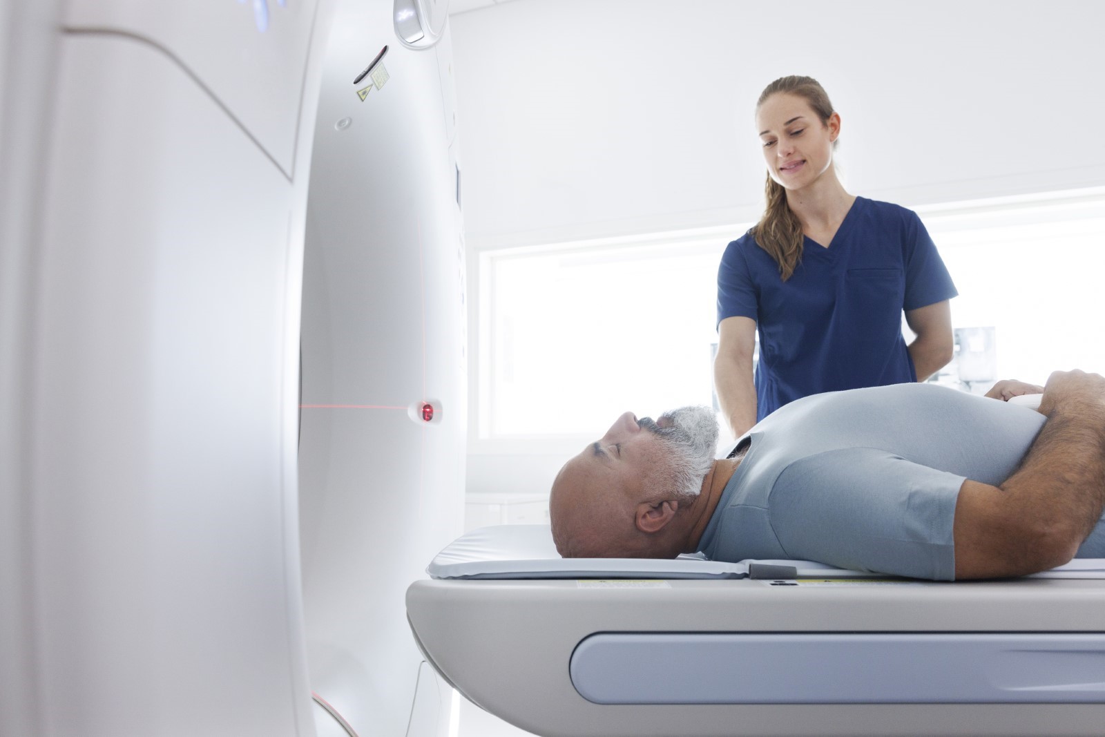
How Do Healthcare Workers Get Exposed to Radiation?
There are three common types of radiation exposure sources:
- The Primary X-ray Beam
- Leakage from the X-ray Tube
- Scattered Radiation
The Primary X-ray Beam
When an X-ray device is activated, X-rays penetrate the body with varying levels of absorption in different tissues based on their radiological density. Typically, physicians and healthcare personnel do not encounter direct exposure to X-ray beams unless their hands or body parts are positioned directly within the irradiation area.
Leakage from the X-ray Tube
Radiation leakage from the X-ray tube is a risk for radiation exposure, especially if the doctor or radiologist operating the machine is physically holding the assembly. If proper precautions are not taken, this can lead to health complications for a medical professional later in life.
Scattered Radiation
Scattered (or backscatter) radiation is the greatest exposure risk for medical professionals. This is radiation that “scatters” into the air as a byproduct of the X-ray beam or tube leakage. As this radiation can be so pervasive in clinics and imaging centers, it is important healthcare professionals take proper precautions.

How Can Doctors Reduce Radiation Exposure to Themselves?
Radiation is an unseen enemy, and this can make it difficult to anticipate its covert long-term damage. But there are many ways doctors can lower radiation exposure:
- Avoid the primary X-ray beam
- Prevent X-ray tube leakage
- Minimize scattered radiation exposure (Time, Shielding, Distance)
- Use ultrasound or MRI instead of CT when possible
- Use augmented reality surgical navigation for procedures
Avoid the Primary X-Ray Beam
Exposure to the X-ray beam itself can lead to cellular damage. Capturing images with collimation and limited magnification are strategies to decrease the intensity of the primary X-ray beam and minimize scattered radiation.
When using fluoroscopy, it is crucial to thoughtfully choose a C-arm fluoroscopy mode, such as pulsed mode or low-dose mode, to prioritize the safety of both the physician and patient in terms of radiation exposure.
Shielding can also assist in radiation protection, which we will discuss more later in the article.
Prevent X-ray Tube Leakage
The X-ray tube may leak radiation. To prevent leakage from the X-ray tube, it is best to install tube shielding. Tube shielding is a material incorporated into the X-ray tube housing to restrict the escape of scattered radiation, safeguarding both patients and staff from exposure. Lead is one of the best materials to use for this function due to its high atomic density.
Minimize Scattered Radiation Exposure
Scattered radiation exposure can be reduced using three strategies: time, shielding, and distance.
Time
Time is the first fundamental safety measure to minimize external radiation exposure. Put simply, the more time a doctor or radiologist spends in close proximity to X-rays or CTs, the more radiation they will be exposed to. By increasing education and skill in the safe use of these machines, healthcare workers can reduce the amount of time they spend close to the radiation source.
In the case of C-arm fluoroscopy, for instance, the doctor must develop their intervention skills to become more efficient, and the radiographer must ensure that the X-ray is checked at the correct location and moment to prevent blurred images.
Shielding
Shielding is the second fundamental safety measure to reduce radiation exposure to healthcare workers. There are many methods of shielding, including shielding clothing, walls, windows, doors, control booths, mobile barriers, thyroid collars, leaded aprons, and leaded safety eyeglasses.
These devices can decrease radiation exposure, but they are only effective if used. Shielding devices are expensive and can be uncomfortable. Therefore, adoption of these devices can be challenging, but their implementation can be paramount.
Distance
Distance is the third safety measure to lessen radiation exposure to doctors. Of the three safety measures, distance may be the most effective when considering the ratio of effort to outcome. Jae Hun Kim may state this best in his Three principles for radiation safety: time, distance, and shielding:
“The amount of [scattered] radiation exposure is not inversely proportional to the distance from the radiation source, but is inversely proportional to the square of the distance. This means that double the distance from the radiation source can reduce the radiation exposure not to 1/2 but to 1/4... In a previous study of radiographers, two steps behind the mobile support structure can decrease the exposure of the radiographer by about 80%. In another study, being only 20 cm farther from the center of the X-ray field can decrease the radiation exposure by about 73%.”
Kim also adds that distance is a free means of protection from radiation exposure. Therefore, all doctors can easily use the principle of increasing distance, where time and shielding are more complex and/or expensive means of protection.
Use Ultrasound or MRI
Modifying the imaging modality used can be another effective way to reduce radiation exposure. Both ultrasound and MRI are non-ionizing forms of radiation, and thus bypass the potential long-term side effects of ionizing imaging modalities.
Ultrasound
Ultrasound, characterized as one of the safest radiology imaging methods, relies on high-frequency sound waves to generate images, without x-rays or ionizing radiation. One significant downside though is that CT scans generally produce higher quality images than ultrasound scans. The reason for this is that ionizing radiation can penetrate the body more sufficiently, thus producing images with greater detail.
Magnetic Resonance Imaging (MRI)
MRI procedures employ magnetic fields and radio waves to capture detailed images of specific body parts, ensuring that there is no associated radiation risk.
Notably, MRI offers superior imaging quality compared to CT scans, especially when healthcare workers require detailed views of soft tissues. The clarity provided by MRI is particularly advantageous in visualizing intricate structures like torn ligaments and herniated discs. This capability makes MRI a preferred option in situations where a comprehensive understanding of soft tissues is crucial for accurate diagnosis and treatment planning.
Despite these advantages, it is important to acknowledge that the cost of MRI procedures for patients is approximately twice that of CT scans. And where a CT may take 1-2 minutes after initial prep, an MRI can take 30-60 minutes. This economic consideration may influence the decision-making process when choosing between imaging modalities. Healthcare providers must weigh the benefits of non-ionizing radiation with the cost of both time and money.
Use AR Surgical Navigation for Procedures
In some circumstances, surgeons use intraoperative fluoroscopy during surgery. This practice exposes both patient and doctor to ionizing radiation, but it is a necessary tool to give surgeons a snapshot view of the inside of the patient. It’s like flipping on an ‘X-ray flashlight’ so they can witness their progress mid-surgery.
Augmented reality (AR) surgical navigation can be a highly effective way to replace intraoperative fluoroscopy. AR surgical navigation combines AR technology with medical imaging to superimpose a 3D image over the body of a patient, so the surgeon can see inside the body without referring to a monitor. So, instead of periodically flipping on an X-ray flashlight during surgery (as is the case with fluoroscopy), it’s like having one on continuously.
Healthcare Workers Striving to Reduce Radiation Exposure
The escalating use of radiological imaging, particularly X-Rays and CT scans, has prompted concerns about the potential risks of radiation exposure to doctors and medical professionals. With a substantial increase in CT scans performed in the U.S., reaching an estimated 70 million annually, and the associated potential for radiation-induced cancers, addressing this issue has become crucial. Despite the undeniable benefits of X-rays and CT scans, the acknowledged risks necessitate the implementation of policies and procedures to minimize radiation exposure for healthcare providers.
Long-term exposure to low-dose radiation poses potential risks, including the development of cataracts, cancer, and cardiovascular diseases. Doctors can employ multiple measures to reduce radiation exposure, including avoiding the primary X-ray beam, preventing leakage from the X-ray tube, and minimizing scattered radiation. Time, shielding, and distance emerge as fundamental safety measures, with the latter proving particularly effective due to its proportional decrease in exposure, as well as its monetary and practical advantages. Additionally, the utilization of ultrasound and MRI, which utilize non-ionizing radiation, presents a safer alternative for specific procedures.
In navigating the balance between the benefits of advanced imaging technologies and the potential risks associated with radiation exposure, healthcare providers play a critical role in implementing and adhering to safety measures. Ultimately, a comprehensive approach that incorporates awareness, education, and the strategic use of innovative technologies can contribute to the goal of minimizing radiation exposure to doctors, while ensuring the continued efficacy of life-saving diagnostic tools.
To significantly reduce the need for intraoperative fluoroscopy, and enhance surgical performance, explore our VisAR augmented reality surgical navigation system. This innovative tool combines radiological imaging and AR technology to produce an immersive surgical “GPS”, enhancing operative accuracy and potential. It is FDA approved for stereotactic spinal surgery and is being used for additional types of surgeries in hospitals around the world.